Digitisation has become an indispensable part of medicine. Nowadays, in certain areas artificial intelligence (AI) can support doctors with the same precision as an expert in this medical field. Medical imaging systems scan the human body, raise alarms in the event of anomalies and can even help to save lives.
With the bodystudio ATBM master, FotoFinder Systems GmbH is now introducing an imaging system that enables physicians to take a new approach to the digital diagnosis of skin cancer. It delivers results in a matter of seconds and can significantly reduce the need for biopsies and excisions.
Since 2013, FotoFinder has focused on the earliest skin cancer diagnosis through Automated Total Body Mapping (ATBM) for fully automated photo documentation of the entire skin surface. Now, the company goes one step further with the new bodystudio ATBM master.
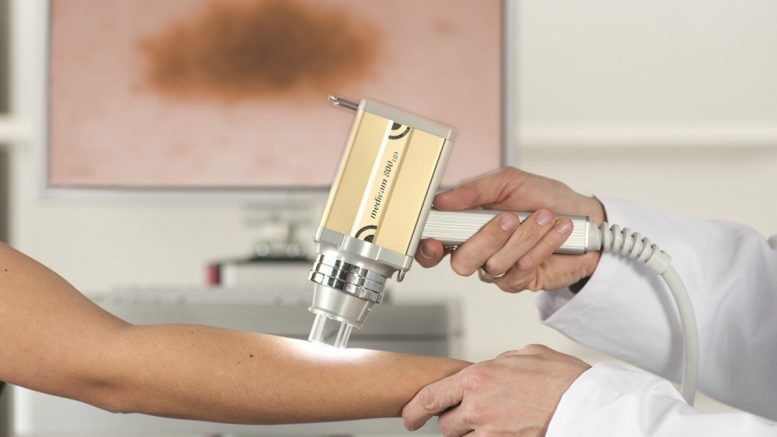
Total Body Dermoscopy is the name of the method by which the entire skin surface is photographed with a special camera and flash system without any reflections and with super-high resolution. This results in excellent clinical images that allow the physician to zoom into the full body photo to such an extent that the microscopic structure of a mole is visible in the overview image. The doctor receives visual support from the fully automatic Bodyscan software, which quickly identifies the existing skin lesions in the whole body image and arranges them according to their relevance. In this way, the physician can quickly “scan” the moles visually without having to examine each one individually using a dermatoscope. Only the few moles that are atypical or suspicious are analysed with the digital dermatoscope. This leads to considerable time savings and enables the detection of even the smallest abnormalities: A fact that can save lives in extreme cases. In any case, the waiting time for the diagnosis and thus also uncertainty and anxiety are considerably shortened. In addition, this method reduces the sometimes painful excision of one or more tissue samples.
“The future of skin cancer diagnostics lies in innovative, intelligent, time-saving solutions,” explains Kathrin Niemela, member of the FotoFinder management board. “Modern analysis methods are largely digital and support physicians in finding abnormalities in many ways.”
Contrary to the widespread opinion, most melanomas do not develop from an existing mole, but appear as new spots, “de novo”, on apparently healthy skin. In most cases, the disease begins with an optically barely perceptible spot, often just 1mm in size, which can however, already contain a malignant cell population of thousands. It is precisely these extremely small lesions that are often overlooked in a classic dermatological examination. Whole-body cartography with the new master technology visualises the moles of a patient in such a way that new lesions become visible at a glance.
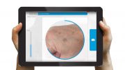
The physician is supported in the analysis and risk assessment of skin lesions by the expert software Moleanalyzer pro, which works with a powerful AI-based deep learning algorithm. According to a clinical study carried out by the Department of Dermatology at Heidelberg University Hospital, AI came up with more accurate diagnostic results than the medical specialists involved in the study – and it only takes the system less than a second each time.*
For patients, the use of Automated Total Body Mapping in combination with AI means greater reliability in the earliest detection of skin cancer. “Compared to the more intuitive approach of a physician, who also includes patient history or genetic predisposition in the diagnosis, the algorithm is absolutely objective in its analysis”, explains Kathrin Niemela from FotoFinder. “The larger, better and more unique the data basis is, the more intelligent the system becomes in a short period of time, thanks to continuous training. Nevertheless, AI cannot replace human intelligence and experience in the detection of skin cancer: In the end, the doctor decides what to do.”


